
Sub-Total: £0.00
Retinal photography has a long and storied history of innovations and innovators, all of whom strove to create a world where proper eye care was readily available to all.
We’ve come a very, very long way since the early days of the fundus eye camera!
In this blog, we’ll run through the history of fundus imaging, present advancements in tech that are already being used to help patients right now, and developments we expect to see in the future of the fundus eye camera.
The Long History of Fundus Imaging
According to research published in the National Center for Biotechnology Information, “Jackman and Webster first published photographs of the human retina in 1886.”
Early fundus photos were limited as a result of poor lighting, overly long exposure times, patient eye movement and prominent corneal reflexes. The results were notably poor, but it was a step in the right direction that sparked a significant development in fundus imaging technology!
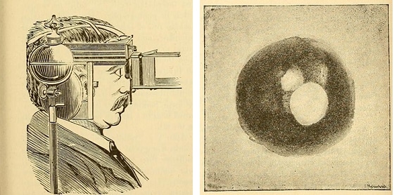
Images from the Ophthalmic Photographers’ Society, illustrations from Jackman and Webster, June 1886.
Following on from these early photographs, “The next breakthrough was the first commercially available fundus camera produced by Carl Zeiss in 1926, following which considerable improvements to the field of view (FoV) were made.”
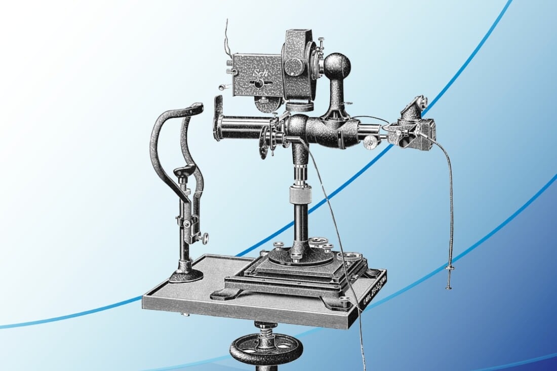
Carl Zeiss’ original fundus eye camera design.
From then, fundus imaging technology started to snowball over the decades, with improvements in optics, film technology and illuminations systems.
Later in the 20th century, cameras started to transition from film-based systems to digital imaging, allowing for quicker image acquisition and easier image storage.
Incremental changes to Zeiss’ pioneering design led us to where we are today, with modern fundus eye cameras able to produce remarkable, high-quality images (ones that Zeiss, Jackman and Webster could only dream of!).
Developments in Modern Fundus Eye Cameras
Fast forward to now; we’ve seen incredible advancements in fundus imaging in the last decade.
From ultra-widefield, full colour confocal imagery through to the use of AI in diagnosing pathologies, our modern fundus cameras are, without a doubt, revolutionary.
The convenience of automation
Automation has become an incredibly important part of modern fundus imaging, too. Traditional joystick-controlled fundus cameras are prone to mistakes, and require a skilled hand (and a still patient!) in order to get the images you need to help with diagnosis.
Instruments such as the iCare DRSplus use an almost ‘hands-free’ system, whereby the technician operating the machine can capture incredibly high-quality images of the rear of a patient’s eye with the touch of a button, allowing for rapid processing and saving optometrists and ophthalmologists significant amounts of time.
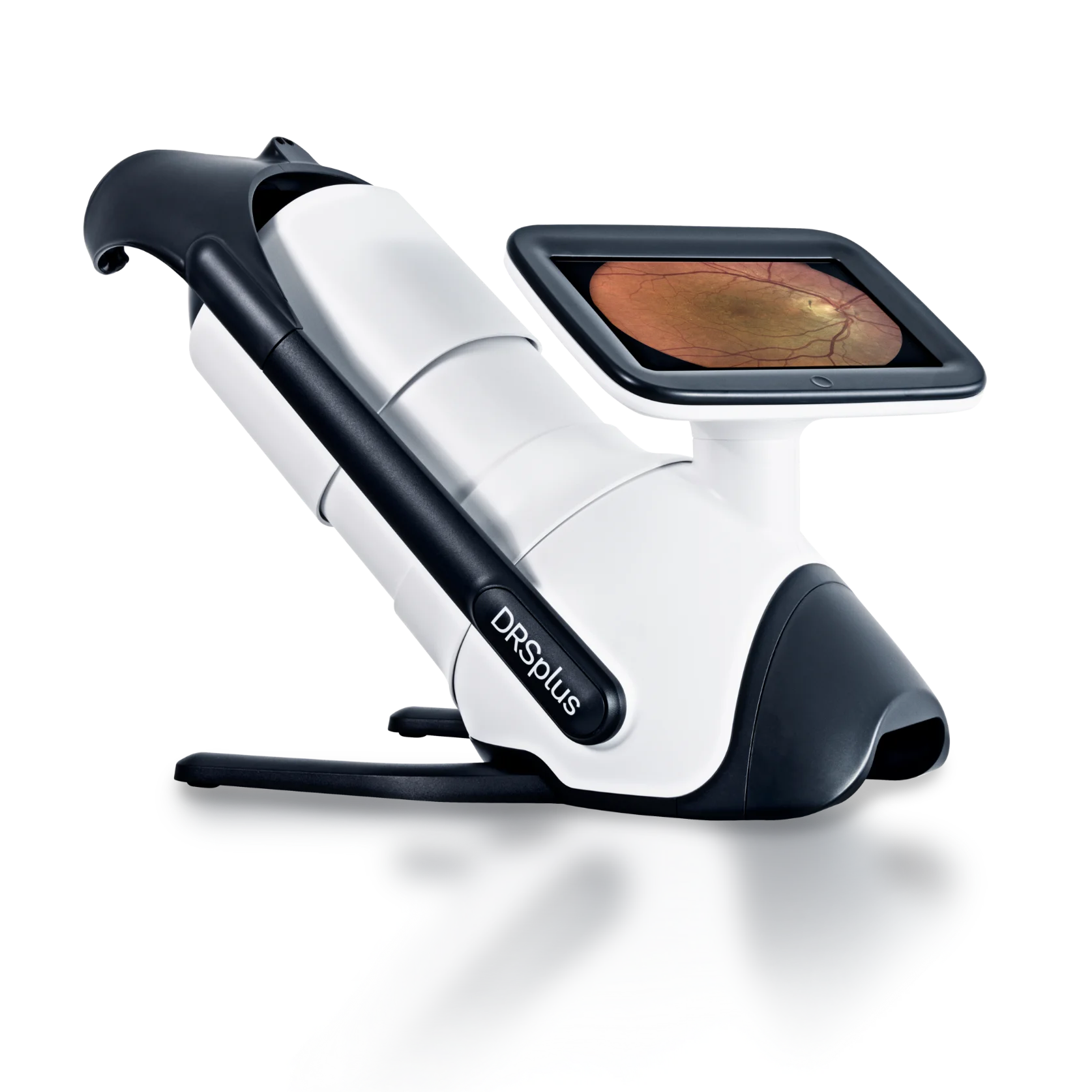
The iCare DRS Plus Fundus Imaging System.
Other modern fundus advancements
As mentioned, we’ve seen both confocal laser technology and AI being used to great effect in modern fundus cameras.
Confocal laser technology is used to reduce or suppress any scattered or reflected light outside the focal plane. With this feature included, the DRSplus guarantees increased image sharpness, better optical resolution and greater contrast when compared to traditional fundus camera imaging.
Advancements in AI are taking much manual analysis out of the ophthalmic diagnostic process, with AI being able to scan imagery and help identify potential issues in an instant. This by no means takes the doctor out of the equation, it simply makes their life easier and more efficient.
AI is being used to dramatically accelerate diagnosis, reduce waiting times, and free up doctors’ time for treatment.
What does the Future of the Fundus Eye Camera Look Like?
Realistically, existing image quality is probably the best that we’re ever going to get; we don’t expect many improvements in this area. We may see some micro improvements here and there, but the image quality we’re currently capable of is the best imaginable (with existing technology). We’re happy to be proven wrong here!
We expect the future of the fundus eye camera to:
Move away from manual controls
What we do expect to happen in the near future is that traditional fundus cameras will become a thing of the past, phased out slowly over years in favour of automated fundus cameras that don’t rely on a joystick. The more human error we can remove from the process, the better!
Combine with other tech
All-in-one instruments are growing in popularity, especially in practices with a finite amount of space.
We’re seeing fundus technology being combined with OCT, but it’s very early days at the moment. Being able to look at both the layers and the surface of the eye simultaneously will be a huge hardware development.
Develop and improve AI usage
We expect to see a huge investment in AI technology, with faster, more elaborate algorithms being able to see potential problems before even the most highly trained doctors would be able to notice them.
As you well know, early diagnosis is paramount in our field, and AI is likely the key in improving how early we’re capable of diagnosing many ailments.
Prioritise ease of use
As discussed earlier, we’re already seeing instruments such as the iCare DRSplus leading the charge in making equipment as easy to use as possible.
This is a trend we’re seeing in modern instruments, and there’s no reason that this trend would cease. The easier ophthalmic tech is to use, the less training staff need to operate, and the more time doctors spend with patients
Dramatically reduce in overall hardware size
Many instruments are already successfully reducing their overall size, such as the handheld iCare IC100 and IC200 tonometers.
In the future, we’ll likely see technologies advancing to the point where portable OCTs and fundus cameras can offer the same imaging quality as our existing equipment. Lightweight, portable ophthalmic tech is one of the keys to ensuring that great eyecare is available to all, even to those in rural, hard-to-reach areas.
Looking to Upgrade Your Fundus Imaging Capabilities?
Then it’s time to talk to our specialists at Mainline!
We work with optometrists and ophthalmologists across the UK, helping you find the perfect instruments that match your practice’s unique needs.
Click the link below to get in touch. A member of our friendly team will be in touch as soon as possible.
Featured products
-
iCare EIDON – Ultra-Widefield Confocal System
Enquire for pricing- Increased field of view up to 200°
- Multiple imaging modalities: TrueColor, blue, red, RedFree, infrared
- Capable of imaging through cataracts and media opacities
- Dilationfree operation through pupils as small as 25 mm
-
iCare DRSplus – TrueColor Confocal Fundus Imaging System
Enquire for pricing- Multiple imaging modalities: redfree, infrared, external, stereo views
- No dilation needed, ensuring comfortable patient experience
- Fast, fully automated imaging through pupils as small 2.5 mm
- Remote Exam feature allows exams to be performed remotely
-
Enquire for pricing
- High-resolution Scheimpflug imaging captures 3D anterior segment
- Contact-free measurement offers rapid, reproducible results
- Detailed corneal and chamber maps enhance diagnostics
- Fast Screening Report simplifies clinical decision layout
-
Enquire for pricing
- 5‑in‑1 system integrates OCT, fundus, biometry, topography
- 80,000 A‑scans/sec delivers rapid, stable 3D imaging
- Single‑shot true‑colour fundus captures clear retinal anatomy
- Detailed OCT‑A visualises microvascular structures without dye
-
Optomed Lumo – Portable Fundus Camera
Enquire for pricing- Non‑mydriatic retinal imaging, undilated pupils
- Large 5″ intuitive touchscreen display
- Wireless DICOM image transfer connectivity
- High‑sensitivity 12 MP sensor optics


-
iCare EIDON – Ultra-Widefield Confocal System
Enquire for pricing- Increased field of view up to 200°
- Multiple imaging modalities: TrueColor, blue, red, RedFree, infrared
- Capable of imaging through cataracts and media opacities
- Dilationfree operation through pupils as small as 25 mm
-
iCare DRSplus – TrueColor Confocal Fundus Imaging System
Enquire for pricing- Multiple imaging modalities: redfree, infrared, external, stereo views
- No dilation needed, ensuring comfortable patient experience
- Fast, fully automated imaging through pupils as small 2.5 mm
- Remote Exam feature allows exams to be performed remotely
-
Enquire for pricing
- High-resolution Scheimpflug imaging captures 3D anterior segment
- Contact-free measurement offers rapid, reproducible results
- Detailed corneal and chamber maps enhance diagnostics
- Fast Screening Report simplifies clinical decision layout
-
Enquire for pricing
- 5‑in‑1 system integrates OCT, fundus, biometry, topography
- 80,000 A‑scans/sec delivers rapid, stable 3D imaging
- Single‑shot true‑colour fundus captures clear retinal anatomy
- Detailed OCT‑A visualises microvascular structures without dye
-
Optomed Lumo – Portable Fundus Camera
Enquire for pricing- Non‑mydriatic retinal imaging, undilated pupils
- Large 5″ intuitive touchscreen display
- Wireless DICOM image transfer connectivity
- High‑sensitivity 12 MP sensor optics
-
TowardPi – BMizar 400KHz Full-Range SS-OCT/A
Enquire for pricing- 400kHz swept-source engine: 400,000 A-scans per second
- Ultra-widefield OCTA: 30 × 25mm single-capture, no stitching
- 10 billion voxels of ultra-high-resolution imaging data
- AI-powered quantification across retina, choroid, and anterior segment
-
Enquire for pricing
- Combines tomography, biometry, refraction, wavefront, retroillumination scanning
- Contact-free axial length measurement for precise biometric data
- Whole-eye wavefront analysis reveals optical aberration profiles
- Supports comprehensive diagnostics for surgery, screening, management
-
Enquire for pricing
- Combined ultra-high-resolution OCT and Scheimpflug imaging
- Single contact-free scan captures whole corneal microstructure
- Aligned data enhances diagnostic accuracy and reproducibility
- Supports advanced corneal assessment and surgical planning
-
iCare EIDON – Ultra-Widefield Confocal System
Enquire for pricing- Increased field of view up to 200°
- Multiple imaging modalities: TrueColor, blue, red, RedFree, infrared
- Capable of imaging through cataracts and media opacities
- Dilationfree operation through pupils as small as 25 mm
-
iCare DRSplus – TrueColor Confocal Fundus Imaging System
Enquire for pricing- Multiple imaging modalities: redfree, infrared, external, stereo views
- No dilation needed, ensuring comfortable patient experience
- Fast, fully automated imaging through pupils as small 2.5 mm
- Remote Exam feature allows exams to be performed remotely


-
TowardPi – BMizar 400KHz Full-Range SS-OCT/A
Enquire for pricing- 400kHz swept-source engine: 400,000 A-scans per second
- Ultra-widefield OCTA: 30 × 25mm single-capture, no stitching
- 10 billion voxels of ultra-high-resolution imaging data
- AI-powered quantification across retina, choroid, and anterior segment
-
Enquire for pricing
- Combines tomography, biometry, refraction, wavefront, retroillumination scanning
- Contact-free axial length measurement for precise biometric data
- Whole-eye wavefront analysis reveals optical aberration profiles
- Supports comprehensive diagnostics for surgery, screening, management
-
Enquire for pricing
- Combined ultra-high-resolution OCT and Scheimpflug imaging
- Single contact-free scan captures whole corneal microstructure
- Aligned data enhances diagnostic accuracy and reproducibility
- Supports advanced corneal assessment and surgical planning
-
iCare EIDON – Ultra-Widefield Confocal System
Enquire for pricing- Increased field of view up to 200°
- Multiple imaging modalities: TrueColor, blue, red, RedFree, infrared
- Capable of imaging through cataracts and media opacities
- Dilationfree operation through pupils as small as 25 mm
-
iCare DRSplus – TrueColor Confocal Fundus Imaging System
Enquire for pricing- Multiple imaging modalities: redfree, infrared, external, stereo views
- No dilation needed, ensuring comfortable patient experience
- Fast, fully automated imaging through pupils as small 2.5 mm
- Remote Exam feature allows exams to be performed remotely
-
£2,995.00 + VAT
- No need for anesthetic drops, air, or calibration
- Automated Measurements Sequence
- Quick IOP readings enhance clinical efficiency
- Simple operation with minimal training required
-
£4,495.00 + VAT
- 200° position freedom
- No need for anesthetic drops, air, or calibration
- Quick Measure for easier and faster IOP measurements
- Enhanced probe control improves handling and measurement precision
-
Enquire for pricing
- Patients measure IOP throughout daily activities independently easily
- Measurements also possible while in supine body position
- Cloud uploads share real world IOP data securely
- Doctors access accurate data supporting better treatment decisions
-
£90.00 + VAT
- Box of 100 probes
- Ensures minimal contact and maximum patient comfort
- Individually packaged for hygienic, contaminationfree measurements
- Simple insertion and removal for efficient workflow
-
iCare ST500 – Slit Lamp Mounted Rebound Tonometer
Enquire for pricing- Slit lamp integration fits most lamps for seamless measurement
- Fast, accurate, and repeatable IOP measurements
- Stable patient positioning for easier measurement
- No need for anesthetic drops, air, or calibration


-
£2,995.00 + VAT
- No need for anesthetic drops, air, or calibration
- Automated Measurements Sequence
- Quick IOP readings enhance clinical efficiency
- Simple operation with minimal training required
-
£4,495.00 + VAT
- 200° position freedom
- No need for anesthetic drops, air, or calibration
- Quick Measure for easier and faster IOP measurements
- Enhanced probe control improves handling and measurement precision
-
Enquire for pricing
- Patients measure IOP throughout daily activities independently easily
- Measurements also possible while in supine body position
- Cloud uploads share real world IOP data securely
- Doctors access accurate data supporting better treatment decisions
-
£90.00 + VAT
- Box of 100 probes
- Ensures minimal contact and maximum patient comfort
- Individually packaged for hygienic, contaminationfree measurements
- Simple insertion and removal for efficient workflow
-
iCare ST500 – Slit Lamp Mounted Rebound Tonometer
Enquire for pricing- Slit lamp integration fits most lamps for seamless measurement
- Fast, accurate, and repeatable IOP measurements
- Stable patient positioning for easier measurement
- No need for anesthetic drops, air, or calibration
-
Enquire for pricing
- Highresolution corneal topography and keratometry
- Noninvasive tear film and dry eye analysis
- Meibography for gland visualisation and assessment
- Contact lens fitting and keratoconus support
-
Enquire for pricing
- Ultracompact slitlamp camera system
- Highresolution photo and video capture
- Swivelling prism for easy operation
- Integrated software with measurements and annotations
-
Tomey MR-6000 Multifunction Unit
Enquire for pricing- All‑in‑one ophthalmic diagnostics for six eye examinations
- Automatic alignment with rapid mode switching workflow
- Integrated dry eye and corneal topography analysis
- Tonometry corrected by pachymetry for accurate IOP
-
£94.50 + VAT
- Precise eyelid retraction and eversion for accurate assessment
- Supports meibography, dry eye, and lid cleaning procedures
- Disposable medicalgrade construction ensures hygiene and patient safety
- Integrated ruler and shaped edges assist clinical examinations efficiently


-
Enquire for pricing
- Highresolution corneal topography and keratometry
- Noninvasive tear film and dry eye analysis
- Meibography for gland visualisation and assessment
- Contact lens fitting and keratoconus support
-
Enquire for pricing
- Ultracompact slitlamp camera system
- Highresolution photo and video capture
- Swivelling prism for easy operation
- Integrated software with measurements and annotations
-
Tomey MR-6000 Multifunction Unit
Enquire for pricing- All‑in‑one ophthalmic diagnostics for six eye examinations
- Automatic alignment with rapid mode switching workflow
- Integrated dry eye and corneal topography analysis
- Tonometry corrected by pachymetry for accurate IOP
-
£94.50 + VAT
- Precise eyelid retraction and eversion for accurate assessment
- Supports meibography, dry eye, and lid cleaning procedures
- Disposable medicalgrade construction ensures hygiene and patient safety
- Integrated ruler and shaped edges assist clinical examinations efficiently
-
Tomey MR-6000 Multifunction Unit
Enquire for pricing- All‑in‑one ophthalmic diagnostics for six eye examinations
- Automatic alignment with rapid mode switching workflow
- Integrated dry eye and corneal topography analysis
- Tonometry corrected by pachymetry for accurate IOP
-
Enquire for pricing
- Compact refraction workstation optimized for efficient eye exams
- Smooth height adjustment supports precise patient alignment
- Integrated storage and cable management for organized setup
- Durable design ideal for busy optometry clinics
-
Huvitz HS-7000/HS-7500 40x Mag Slit Lamp
Enquire for pricing- Five‑step magnification drum provides versatile visual detail
- Galilean optics enhance clarity for accurate clinical diagnosis
- Multiple built‑in filters improve contrast for specific tests
- Joystick with trigger simplifies positioning and image capture
-
Huvitz HDR-9000 Digital Refractor
Enquire for pricing- 21‑Point refraction method ensures systematic exam precision
- Wireless Wi‑Fi communication connects to clinical devices
- Dual cross‑cylinder lenses support accurate astigmatic testing
- Tiltable body and height control enhance patient comfort


-
Tomey MR-6000 Multifunction Unit
Enquire for pricing- All‑in‑one ophthalmic diagnostics for six eye examinations
- Automatic alignment with rapid mode switching workflow
- Integrated dry eye and corneal topography analysis
- Tonometry corrected by pachymetry for accurate IOP
-
Enquire for pricing
- Compact refraction workstation optimized for efficient eye exams
- Smooth height adjustment supports precise patient alignment
- Integrated storage and cable management for organized setup
- Durable design ideal for busy optometry clinics
-
Huvitz HS-7000/HS-7500 40x Mag Slit Lamp
Enquire for pricing- Five‑step magnification drum provides versatile visual detail
- Galilean optics enhance clarity for accurate clinical diagnosis
- Multiple built‑in filters improve contrast for specific tests
- Joystick with trigger simplifies positioning and image capture
-
Huvitz HDR-9000 Digital Refractor
Enquire for pricing- 21‑Point refraction method ensures systematic exam precision
- Wireless Wi‑Fi communication connects to clinical devices
- Dual cross‑cylinder lenses support accurate astigmatic testing
- Tiltable body and height control enhance patient comfort
-
Enquire for pricing
- Measures refraction, axial length, keratometry in one
- Software guides complete myopia assessment and reporting process
- Quick, non-contact measurement quality supports painless exams
- Reliable results help detect myopia progression early
-
Enquire for pricing
- Combines tomography, biometry, refraction, wavefront, retroillumination scanning
- Contact-free axial length measurement for precise biometric data
- Whole-eye wavefront analysis reveals optical aberration profiles
- Supports comprehensive diagnostics for surgery, screening, management

-
Enquire for pricing
- Measures refraction, axial length, keratometry in one
- Software guides complete myopia assessment and reporting process
- Quick, non-contact measurement quality supports painless exams
- Reliable results help detect myopia progression early
-
Enquire for pricing
- Combines tomography, biometry, refraction, wavefront, retroillumination scanning
- Contact-free axial length measurement for precise biometric data
- Whole-eye wavefront analysis reveals optical aberration profiles
- Supports comprehensive diagnostics for surgery, screening, management


