
Uveitis is when an inflammatory disease produces swelling and redness within the eye, and destroys the eye tissue. If left untreated it can lead to scarring or vision loss.
The inflammation occurs within the uvea, which is the middle layer of the eye, which includes the iris, ciliary body and choroid. However, it can also affect the lens, retina, optic nerve and vitreous.[1]
There are 4 types which are[2]:
- Anterior uveitis – This is the most common form. It occurs when the iris and the ciliary body become inflamed, and has been linked rheumatologic, skin, gastrointestinal, lung and infectious diseases.
- Intermediate uveitis – This occurs most commonly in children and young people, when the area behind the ciliary body and the retina becomes inflamed. This type has been linked to sarcoidosis and multiple sclerosis.
- Posterior uveitis – This is the least common type. It occurs when the choroid and the retina at the back of the eye becomes inflamed and is often called choroiditis or chorioretinitis.
- Panuveitis uveitis – This is when all 3 parts of the uvea are affected by inflammation.
Treatment is done with steroid medicine to reduce the eye inflammation. These can be eyedrops, injections, tablets or capsules but in extreme cases surgery may be required[3].
Detection
Eye Pressure – An Ophthalmologist could use a tonometer to test the eyes IOP to gage inflammation and swelling.
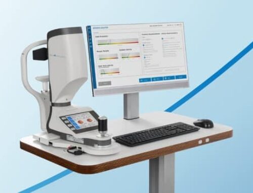
OCT Scan – Optometrists could use OCT scans to more accurately detect and manage the issue. By providing a clearer image, OCT scans allow them to detect certain signs and complications of the different types by showing details of each of the eyes layers.
To see our full range of tonometers, click here. Or to see our full range of OCT’s click here.
References:
[1] https://www.specsavers.co.uk/eye-health/oct-scan/conditions/detecting-uveitis-symptoms
[2] https://www.lei.org.au/services/eye-health-information/uveitis/
[3] https://www.nhs.uk/conditions/uveitis/


