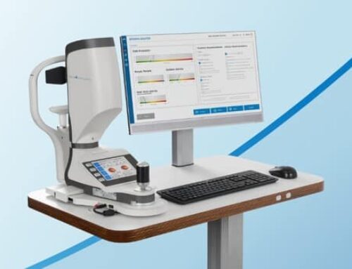
Optical coherence tomography (commonly referred to as OCT) is a non-invasive imaging test. It uses light waves to take cross-section pictures of the retina, and lens if an anterior segment module is used.
With OCT, an ophthalmologist can see the retina’s different layers. This allows the ophthalmologist to measure their thickness and see anomalies. The equipment will scan the eye without touching it.
OCT is can be used in diagnosing eye conditions, such as[1]:
- macular hole
- macular pucker
- macular edema
- age-related macular degeneration
- glaucoma
- central serous retinopathy
- diabetic retinopathy
- vitreous traction
In addition, Optical Coherence Tomography is often used to evaluate disorders of the optic nerve. The OCT exam helps the ophthalmologist see changes to the fibers. This can be used when diagnosing glaucoma.
You can see our range of anterior segment OCTs and retinal OCTs on our product page.
References
[1] https://www.aao.org/eye-health/treatments/what-does-optical-coherence-tomography-diagnose


