Jonathan Sugden

HomeJonathan Sugden

About Jonathan Sugden

After gaining a first-class honours degree in commercial photography at the University of Derby, Jonathan became an ophthalmic photographer for the NHS in Doncaster. He would perform a range of ophthalmic imaging examinations every day, such as OCT, ultrasound and photography. Over the last 6 years, he has worked within the industry helping customers to select the right equipment for their needs and supporting them with training and support after installation.
17 08, 2021

The DRSPlus: Positive customer feedback

2023-02-14T16:06:00+00:00August 17th, 2021|News|

Customer feedback allows us to understand the real-world challenges faced by the market we serve. That’s why we make a point of asking for product feedback every time a customer purchases or trials one of the devices we offer.

When it comes to the iCare DRSplus Fundus Imaging System, the feedback is always positive and really showcases the performance and ease-of-use […]

13 08, 2021

Sunderland Eye Infirmary Trial DRSPlus

2023-02-14T15:50:22+00:00August 13th, 2021|News|

For a busy eye emergency department like Sunderland Eye Infirmary, it can be difficult to find the time to trial and upgrade existing ophthalmic equipment. To support this busy team of doctors and specialists in finding fundus imaging equipment that could improve patient comfort and diagnostic accuracy, Mainline Instruments worked with Ophthalmic Photographer, Hugh Harris to complete a comprehensive trial […]

1 07, 2021

Five key features of the DRSplus

2021-08-13T11:14:28+01:00July 1st, 2021|News|

The DRSplus device from iCare sets a class-leading standard in retinal imaging thanks to its confocal fundus imaging system and white LED illumination.

Compared to other market-leading alternatives, the state-of-the-art DRSplus is a feature-rich fundus imaging camera that offers many impressive plus points for both large chain and smaller independent opticians.

Here, we explore five key features of the iCare DRSplus:


Multiple imaging […]

4 05, 2021

What is age-related macular degeneration?

2021-07-23T14:02:16+01:00May 4th, 2021|News|

Age-related macular degeneration (AMD) is caused by the deterioration of the central area of the retina, that sends images via the optic nerve from the eye to the brain. The retina’s central portion, known as the macula, is responsible for central vision in the eye, and it controls our ability to read, drive a car, recognise faces or colours, and […]

16 04, 2021

Who invented the ophthalmoscope?

2021-07-23T14:01:43+01:00April 16th, 2021|News|

An ophthalmoscope is a device used for examining the interior structures of the eye, including the fundus and retina.

It was invented in 1851 by Hermann Von Helmholtz. He was a professor of anatomy and physiology in Königsberg, Germany.[1]

He wanted to show his students the emission of reflected light from the eye. The first model was constructed of pasteboard, […]

16 04, 2021

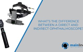
What is the difference between a direct and indirect ophthalmoscope?

2023-12-19T14:34:27+00:00April 16th, 2021|News|

Ophthalmoscopy is a test that look at the back of the eye called the fundus. The fundus consists of the retina, optic disc and blood vessels.[1]

The test is usually included in a routine eye exam. It is also necessary to perform ophthalmoscopy if you have a condition that affects your blood vessels, which can include high blood pressure […]

31 03, 2021

How can eyes be affected by a brain injury?

2023-02-14T16:42:31+00:00March 31st, 2021|News|

Eye and vision problems can be a fairly common occurrence after a head or brain injury. Some vision problems can be due to trauma to the eye, but many other vision problems are actually caused by an injury to the brain.

Even mild head injuries, such as whiplash from a car incident, can cause vision problems. Examples of this could be […]

23 03, 2021

What features should your fundus equipment have in 2021?

2023-02-14T16:21:13+00:00March 23rd, 2021|News|

Fundus photography is a vital component in ophthalmology and optometry. Detection and documentation of the posterior eye is crucial in disease management. The fundus imaging technology used to view the ophthalmoscopic appearance of a patient’s retina is ever-changing, so what features should your fundus imaging equipment have in 2021[1]?

1) Whilst most fundus cameras are non-mydriatic (can […]

12 03, 2021

What Is Optical Coherence Tomography (OCT)?

2023-02-14T16:19:55+00:00March 12th, 2021|News|

Optical coherence tomography (commonly referred to as OCT) is a non-invasive imaging test. It uses light waves to take cross-section pictures of the retina, and lens if an anterior segment module is used.


With OCT, an ophthalmologist can see the retina’s different layers. This allows the ophthalmologist to measure their thickness and see anomalies. The equipment will scan the eye without […]